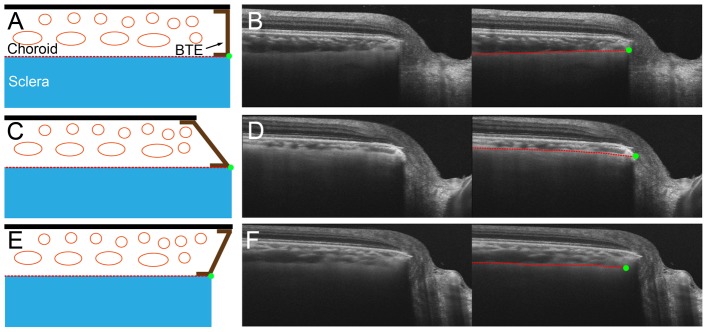
Figure 2. Schematic diagram and sample images for detecting the anterior scleral canal opening (ASCO) in various types of border tissue of Elschnig (BTE).

(A,B) non-oblique BTE, (C,D) externally oblique BTE, (E,F) internally oblique BTE. B, D, F. Left column is SS-OCT image without label, and right column is SS-OCT image with label. Anterior scleral surface (red dashed lines) is followed to the optic nerve head, and projected to define the ASCO (green dots).
