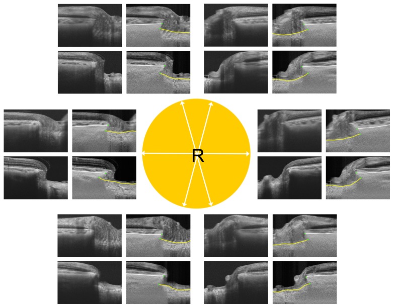
Figure 4. Comparison of ALI positions between eyes from a patient with primary open-angle glaucoma (POAG) and a normal control (both aged 62 years).
The images are for the right eye in both patients. White arrows in the central circle indicate the meridians of the scans. The upper and lower images in each pair are from the normal control and POAG eyes, respectively. Yellow lines indicate the lamina cribrosa surface and white lines indicate the choroidoscleral interface. Note that ALID is greater in the POAG eye, with the most prominent differences being observed in the superotemporal and inferotemporal areas.

