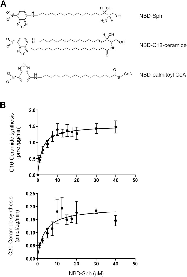
Fig. 1.
Structure of NBD-lipids and optimization of CerS assay using NBD-Sph. A: Structure of NBD-lipids. B: Homogenates from cells overexpressing CerS5 or CerS4 were assayed in a 20 μl reaction volume with increasing amounts of NBD-Sph with 50 μM C16-CoA or C20-CoA, respectively, and 20 μM defatted BSA, at 37°C. Results are means ± SEM of two to three independent experiments.

