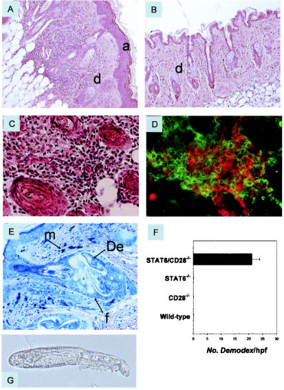
FIG. 3.
Dermatitis associated with Demodex infestation in STAT6/CD28−/− mice. (A) Hematoxylin- and eosin-stained skin section (magnification, ×200) of an 8-month-old STAT6/CD28−/− mouse revealed acanthosis and a dense lymphoid infiltrate in the dermis extending into the subcutaneous fat. In contrast, skin sections from age-matched STAT6−/− mice (B), CD28−/− mice (data not shown), and WT mice were comparable, and there was no indication of dermatitis. (C and D) High magnification (×400) of the deep dermis of STAT6/CD28−/− mice revealed extensive lymphoid infiltration (C) composed of both CD4 (red) and CD8 (green) T cells (D). (E) Giemsa staining (magnification, ×400) of hair follicles in the skin of 3-month-old STAT/CD28−/− mice revealed numerous Demodex ectoparasites and increased numbers of mast cells. (F) Frequency of Demodex mites in randomly selected high-power fields (magnification, ×200) of hematoxylin- and eosin-stained skin sections (four mice per treatment group) from age-matched STAT6/CD28−/−, STAT6−/−, CD28−/−, and WT mice. (G) Isolated ectoparasites (magnification, ×400) identified as D. musculi. a, acanthosis; d, dermis; ly, lymphoid infiltrate; m, mast cells; De, Demodex; f, follicle; hpf, high-power field.

