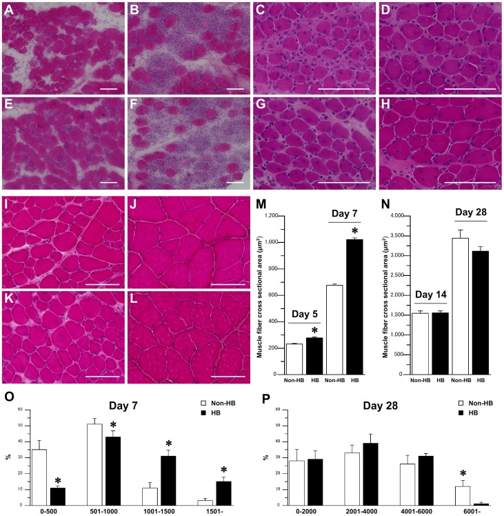
Figure 1. Skeletal muscle regeneration after injury.
Representative muscle fiber cross-sections stained with hematoxylin and eosin in the non-hyperbaric (Non-HB; A–D, I, J) and hyperbaric (HB; E–H, K, L) groups at 24 (A, E) and 48 (B, F) h, and 5 (C, G), 7 (D, H), 14 (I, K), and 28 (J, L) days after injury. Bar = 100 µm. Cross-sectional area of centrally nucleated muscle fibers 5 and 7 days (M), and 14 and 28 days (N) after injury. Distribution of centrally nucleated muscle fiber cross-sectional area at 7 (O) and 28 (P) days after injury. Three sections per animal and 3 randomly chosen fields per section were evaluated. In each section, over 50 centrally nucleated muscle fibers were measured. Values represent means ± standard error (SE). * is significantly different from the Non-HB group, p<0.05.

