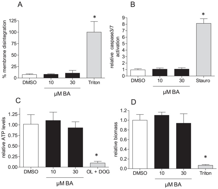
Figure 2. Cytotoxicity of BA in MEF.
MEF were treated with BA (10 µM and 30 µM) for 48 h before they were subjected to determination of membrane integrity (% LDH release) (A), potential of proapoptotic events (activation of caspase 3/7) (B), of ATP levels (C) and biomass (D). Bar graphs depict compilation of three independent experiments (expressed as fold of the DMSO mean value in B, C, and D), each in quadruplicate (mean + SD, * p<0.05, ANOVA, Dunnett's post test versus DMSO ctrl). Staurosporine (Stauro, 1 µM for 6 h), triton (1% for 1 h) or a combination of oligomycin A (OL; 2 µM) and DOG (10 mM; 5 h) served as positive controls in the assays.

