Figure 1.
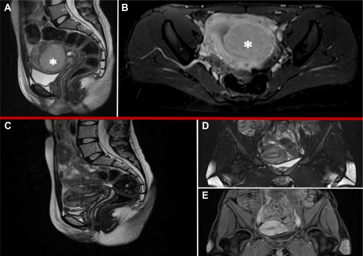
MRI features: preoperative (A, B) and 6 months after surgery (C–E).
Notes: (A) Saggital-T2 weighted imaging of well-defined uterine lesion with dishomogeneous signal intensity. (B) Axial-T2 weighted imaging with contrast enhancement showing early intense strengthening in the postenhancement phase. (C) Saggital-T2 weighted imaging of regular uterus. (D) Coronal-T2 weighted imaging of regular uterus. (E) Coronal-T1 weighted imaging with low-contrast enhancement in area of previous surgery. *indicates the uterine lesion.
Abbreviation: MRI, magnetic resonance imaging.

