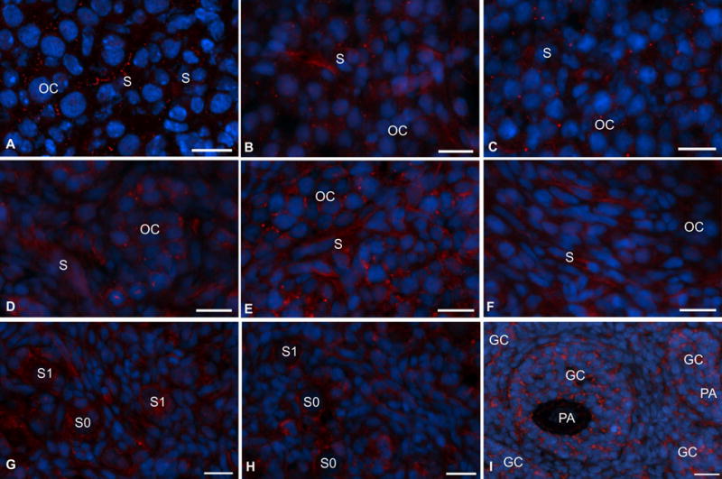
Fig. 5.

Immunofluorescence localization of FSHR protein in hamster ovaries during perinatal development. (A) embryonic age 14 (E14). (B) E15, (C) postnatal day 1 (P1), (D) P2, (E) P4, (F) P6, (G) P9, (H) P10 and (I) P14. Scant punctate red (FSHR) fluorescence was visible in the oocyte cluster (OC) as well as somatic cells (S) as early as E14. Oc, oocyte clusters, S, somatic cells, S0, primordial follicles, S1, primary follicles, GC, granulosa cells, PA, preantral follicles. FSHR= red, Nuclei= blue. Bar = 10 μm.
