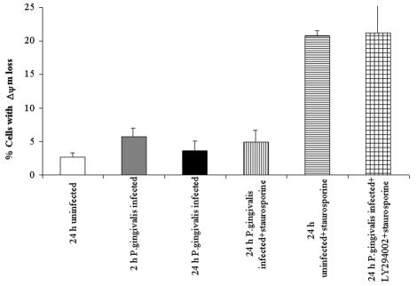
FIG. 5.
Analysis of ΔΨm in P. gingivalis infection. The conditions were similar to those for Fig. 4. Twenty-four-hour-infected and uninfected (control) cells were incubated with or without LY294002 for 18 h before treatment with staurosporine for 3 h (21 h postinfection). Samples (24 h postinfection) were stained with DiOC6 (40 nM) to measure mitochondrial ΔΨm and analyzed by flow cytometry. The bars represent the percentages of cells with loss of ΔΨm. Data are expressed as means from at least two separate experiments, and the values represent the means and standard deviations of separate measurements.

