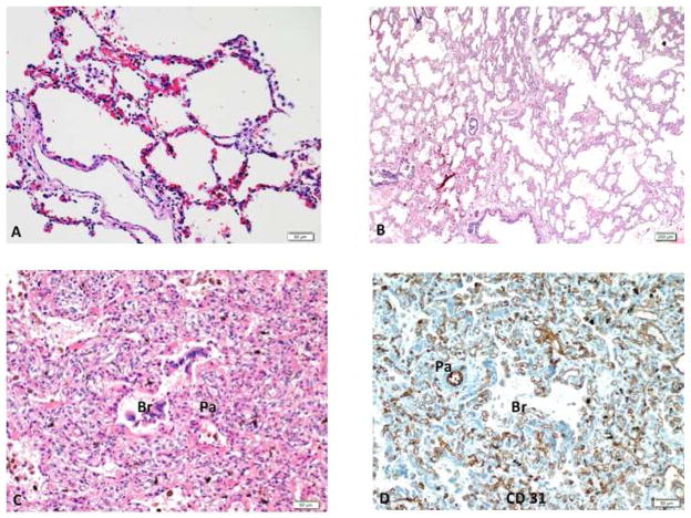
Figure 1.
Lung histology from an infant with congenital diaphragmatic hernia shows 3 distinct histologic patterns. The BPD like is characterized by enlarged and simplified alveoli identical to that seen in infants with BPD (A). The immature pattern demonstrates lung tissue composed of immature alveoli with thickened interstitium (B). A “hemangiomatosis” pattern is dominated by numerous capillaries filling up the interstitium, as well as surrounding the airways (Br) and pulmonary artery (PA) (C), highlighted by CD31 immunostaining (D). BPD – bronchopulmonary dysplasia.

