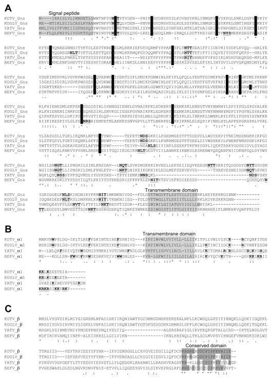
Figure 2.
Clustal X alignments of the deduced amino acid sequences of KOTV, KOOLV, YATV and BEFV accessory proteins. A. GNS proteins showing predicted signal peptide (SignalP) and transmembrane (TMHMM) domains (shaded in grey), cysteine residues in the ectodomain (shaded in black) and potential N-glycosylation sites (bold and underlined). B. α1 proteins showing characteristics of Class I viroporins including predicted transmembrane domains (shaded in grey), large aromatic residues in the N-terminal domain and basic residues in the C-terminal domain (bold and underlined). C. β proteins showing the conserved domain near the C-terminus (shaded).

