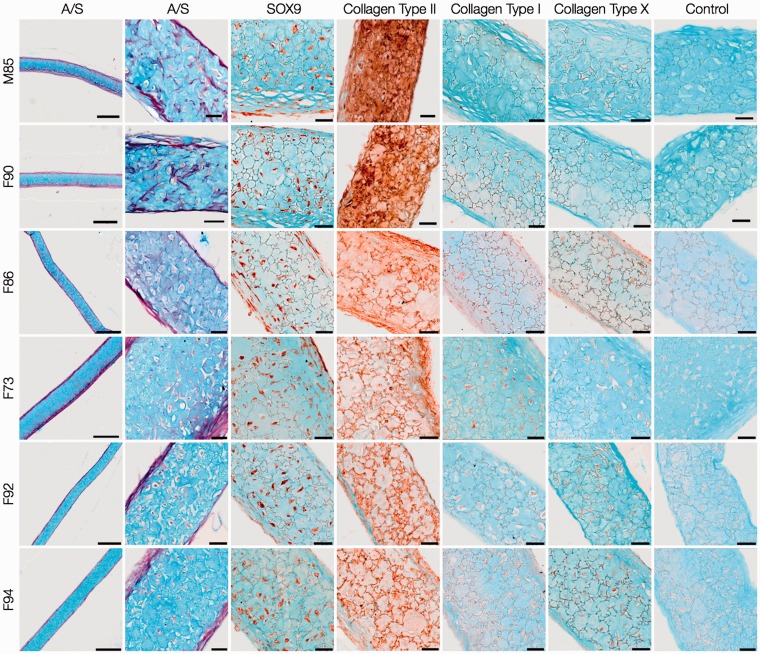
Figure 6.
Histological and immunohistochemical analysis of day-28 HAC explants. HACs from six osteoarthritic individuals (M85, F90, F86, F73, F92, F94) were cultured in Alvetex® 3-D scaffolds for 28 days in chondrogenic media. Sections of the cartilaginous explants demonstrated presence of chondrocytes in lacunae embedded in Alcian blue-stained proteoglycan matrix surrounded by a thin layer of Sirius red-stained fibrous collagen along the periphery. Chondrogenic differentiation was confirmed by robust staining for SOX-9 and collagen Type II, and negligible staining for collagens Type I and Type X. Scale bars for low and high magnification images represent 200 µm and 50 µm, respectively.

