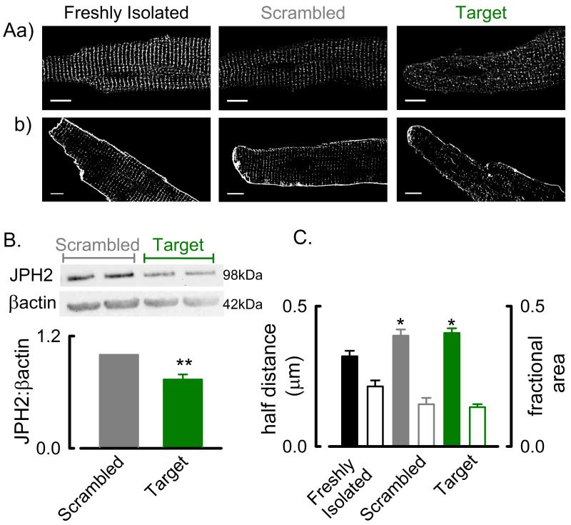
Figure 7. Junctophilin 2 gene silencing does not reduce t-tubule density in rat ventricular cells.
A. Representative images showing immuno-localisation of JPH2 (a) and membrane staining with di-4-ANEPPS (b) in freshly isolated (left), scrambled siRNA treated (centre) and JPH2 siRNA treated (right) rat ventricular cells. B. Example Western blots of JPH2 and β-actin in scrambled and JPH2 target treated rat ventricular cells (upper) and mean data showing reduced JPH2 protein abundance in JPH2 gene silenced cells (lower). **, P < 0.01. N = 4 experiments. C. Mean data showing no change in half-distance (solid bars) or fractional t-tubule area (open bars) measurements following JPH2 gene silencing in adult rat ventricular myocytes. *, P < 0.05 vs freshly isolated myocytes. Data from (cells/experiments); freshly isolated, 20/5; scrambled, 48/5; target, 51/5.

