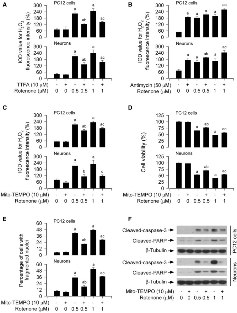
FIG. 7.
Mitochondrial H2O2 elicits apoptosis in rotenone-exposed neuronal cells. The indicated cells were treated with (A and B) rotenone (0.5 and 1 μM) in the presence or absence of TTFA (10 μM) or antimycin A (50 μM) for 24 h, or (C, D, E, and F) pretreated with/without Mito-TEMPO (10 μM) for 1 h and then exposed to rotenone (0.5 and 1 μM) for 24 h, followed by (A, B, and C) H2O2 imaging using a peroxide-selective probe H2DCFDA, (D) cell viability evaluation using the MTS assay, (E) cell apoptosis analysis using DAPI staining, or (F) Western blotting using the indicated antibodies. For (F), the blots were probed for β-tubulin as a loading control. Similar results were observed in at least three independent experiments. For (A), (B), (C), (D), and (E), all data were expressed as means ± SEM (n = 6). ap < .05, difference with control group; b p < .05, difference with 0.5 μM rotenone group; cp < .05, difference with 1 μM rotenone group.

