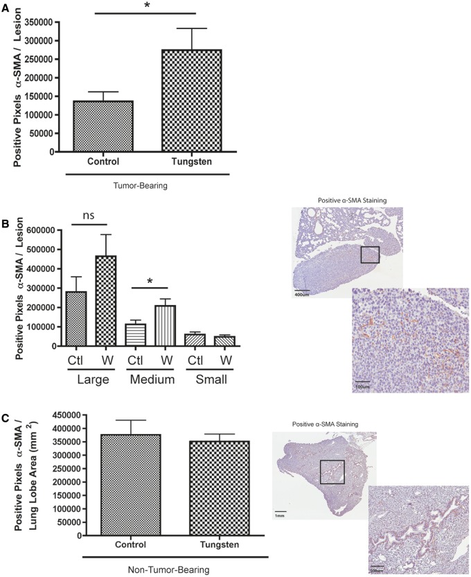
FIG. 5.
Tungsten increases the α-SMA positive, cancer-associated fibroblasts at the metastatic site. A, Number of α-SMA positive pixels per metastatic lesion in the lung. Graph shows mean ±SE for control and tungsten-exposed, metastatic lesions. *P < 0.05 unpaired Student’s t test. B, Number of α-SMA positive pixels per metastatic lesion stratified by lesion size (C = control, W = tungsten). *P < 0.05 unpaired Student’s t test per stratified pair. Representative images show positive α-SMA staining in metastatic lesions (6× and 20×). C, Number of α-SMA positive pixels per lobe in non-tumor-bearing mice lung tissue. Graph shows mean ± SE for control and tungsten-exposed, non-tumor-bearing animals (n = 8 lung lobes). Representative images show positive α-SMA staining in normal lung tissue (2 × and 4×).

