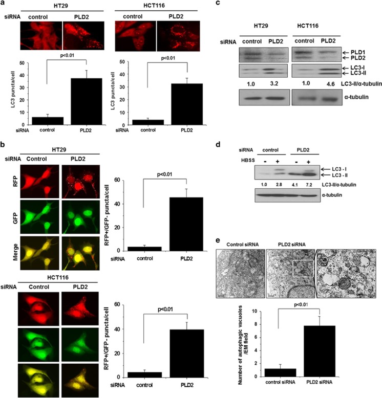
Figure 1.
Depletion of PLD2 induces autophagy. (a) HT29 and HCT116 cells were transfected with an siRNA directed against PLD2 and then immunostained with antibody specific for LC3. Endogenous LC3 punctate dots were observed by fluorescence microscopy, and the number of puncta per cell was quantified (7–10 cells were assessed). The data are representative of three independent experiments. (b) The cells were co-transfected with mRFP-GFP-LC3 and a PLD2-directed siRNA for 24 h, and the total number of RFP-positive/GFP-negative puncta per cell was counted. The data are representative of three independent experiments. (c) The cells were transfected with an siRNA directed against PLD2, and the lysates were immunoblotted with the indicated antibodies. The levels of LC3-II compared with that of α-tubulin were quantified using densitometer analysis. (d) HT29 cells were transfected with an siRNA directed against PLD2 and then cultured under amino acid and serum starvation conditions (HBSS media) for 6 h, after which the lysates were immunoblotted with the indicated antibodies. The levels of LC3-II compared with that of α-tubulin were quantified by densitometer analysis. (e) HT29 cells were transfected with the indicated siRNAs and then fixed and examined by transmission electron microscopy to detect autophagic vacuoles, which were counted in the field of view. Arrows indicate autophagic vacuoles. The values are the means±s.d. of three independent experiments.

