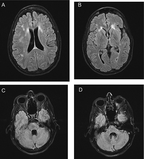
Figure 2.

Fluid-attenuated inversion recovery - conventional spin echo sequence images of the patient’s brain at two-year follow-up. Lesions persist in the periventricular areas and white matter (A,B), but show decreased signal in the pons (C) and middle cerebellar peduncle (D) relative to corresponding images in Figure 1.
