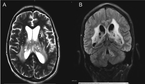
Figure 1.

Brain magnetic resonance image: axial and coronal views showing clusters of cystic structures within the lateral ventricles periventricular white matter area and basal ganglia. There is enlargement of the ventricular system as well. B) shows additional cystic lesions surrounding the fourth ventricle.
