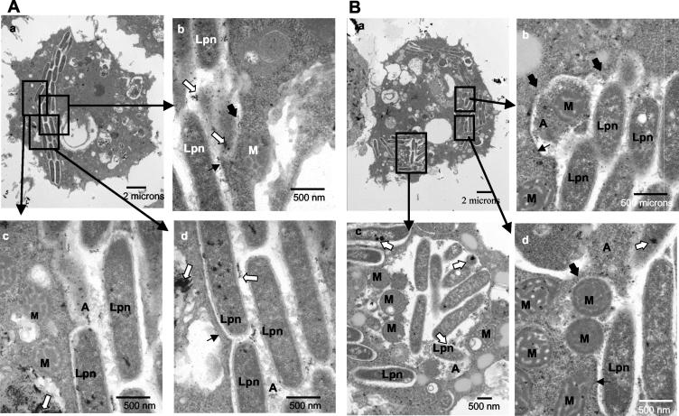
FIG. 5.
Presence of lysosomal contents in the LCP in A. polyphaga at 12 h. Shown are representative electron micrographs of L. pneumophila-infected A. polyphaga at 12 h postinfection where the bacteria are in a disrupted phagosome (A) or cytoplasmic (B). Thick black arrows show sites without any visible phagosomal membrane. Thin black arrows show sites where the phagosomal membrane is still visible. Lysosomal acid phosphatase is indicated by the large white arrows. In panel A, the phagosomal membrane is disrupted (large black arrows) and amorphous material (A) is present within the phagosome. In panel B, although parts of the phagosomal membrane are still visible (thin black arrows), L. pneumophila (Lpn) bacteria are mostly cytoplasmic, as shown by the presence of amorphous material (A) and the presence of mitochondria (M) dispersed among the bacteria.

