Figure 3.

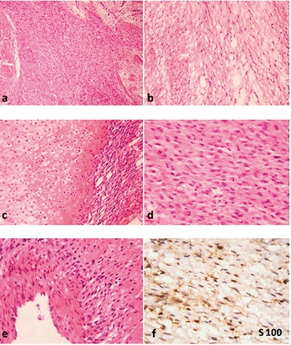
a) Cellular spindle cell tumor with focal myxoid areas; b) myxoid areas with serpentine cells; c) cartilaginous differentiation; d) high mitotic activity in tumor cells; e) plump tumor cells around blood vessel; f) focal S100 positivity.

a) Cellular spindle cell tumor with focal myxoid areas; b) myxoid areas with serpentine cells; c) cartilaginous differentiation; d) high mitotic activity in tumor cells; e) plump tumor cells around blood vessel; f) focal S100 positivity.