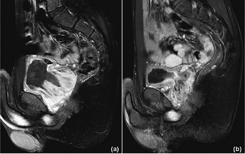
Figure 2.

Prostatic stromal sarcoma: magnetic resonance imaging at diagnosis (a), and after 3 cycles of chemotherapy (b) (T1-fat-suppressed sequences with contrast enhancement). a) Non-homogeneous, vascularized mass (6×6×6.5 cm) originating from the prostate and extending into the bladder and seminal vesicles with signs of rectal wall infiltration. b) Significant shrinkage is evident (2×2×6.5 cm), with an estimated volume reduction of approximately 85%.
