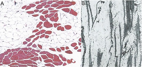
Figure 1.

A) Histological appearance of the infiltrative intramuscular lipoma. Mass of mature uni-vacuolated adipocytes of fairly uniform size, which irregularly infiltrate between muscle fibers. Transverse section showing chequerboard-like appearance. Reproduced with permission (D’Alfonso™, 2011; Copyright 2011 College of American Pathologists).37 B) Histological appearance of the infiltrative intramuscular lipoma. Mass of mature uni-vacuolated adipocytes of fairly uniform size, which irregularly infiltrate between muscle fibers. Longitudinal section showing the striated appearance of the muscle fibers caused by the proliferation of fat cells. Reproduced with permission (Kindblom LG, 1974; Copyright 1974 American Cancer Society).27
