. 2014 Sep 11;7(1):60–68. doi: 10.4168/aair.2015.7.1.60

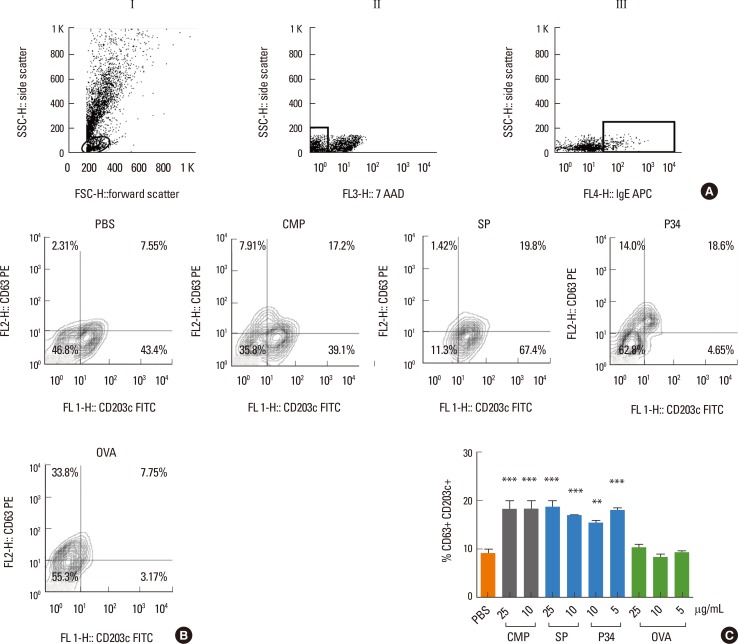
Fig. 2. Basophil activation test by flow cytometry. Basophils from healthy donor subjects were stripped and passively sensitized with milk-specific IgE-containing sera from milk-allergic patients (n=5). (A) Cells were selected according to physical parameters (AI), live cells (AII) and IgE-membrane-bound cells (AIII). (B) Double-positive cells expressing CD63 and CD203c were analyzed in the later gate when different allergens at various concentrations were used to induce basophil activation. Results corresponding to a representative experiment are shown. (C) Percentages of CD63+ CD203c+ cells with all positive sera analyzed. Data represent means±SEM % of double-positive basophils. Statistically significant difference by ANOVA: *P<0.05 , **P<0.01, ***P<0.005, vs PBS group. CMP, cow's milk proteins; SP, soy proteins; OVA, ovalbumin.

Fig. 2