Figure 10.

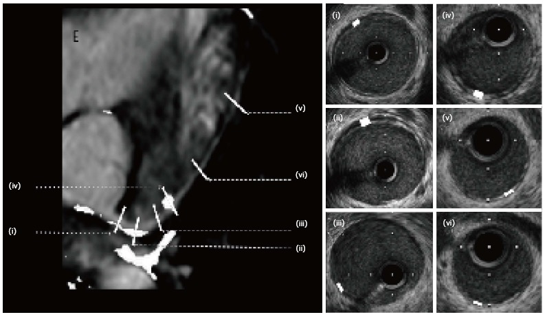
Late gadolinium enhancement in the coronary vessel wall showing corresponding positions for intravascular ultrasound: Illustrates intimal thickening corresponding to enhancement on overlay picture on the left.

Late gadolinium enhancement in the coronary vessel wall showing corresponding positions for intravascular ultrasound: Illustrates intimal thickening corresponding to enhancement on overlay picture on the left.