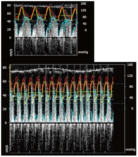
Figure 7.

(A) Resting pressure and flow recording (Red: Aortic pressure; Yellow: Distal coronary pressure; Blue: Pulse wave Doppler envelope) and (B) during hyperemia note that the aortic pressure has decrease as well as the distal coronary pressure. FFR: Ratio of the mean distal coronary pressure at a point past the stenosis the aortic pressure during maximal hyperemia; CFR: Ratio of hyperemic blood flow to resting myocardial blood flow.
