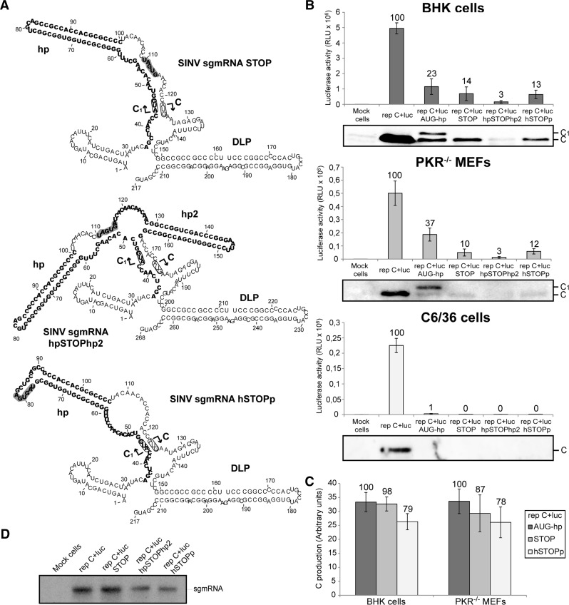
FIGURE 10.
Translation of SINV sgmRNAs bearing termination codons at different positions. (A) RNA secondary structure of 5′ UTRs predicted by RNAfold (see legend in Fig. 1A). (B) BHK (upper panel), PKR−/− MEFs (middle panel), and C6/36 (lower panel) cells were transfected with Lipofectamine 2000 and the corresponding SINV replicons transcribed in vitro. Seven, 5, and 8 h later, respectively, cells were harvested to measure luciferase activity. Values are plotted as means ± SD of three independent experiments. The percentage values obtained from mutant replicons relative to control rep C+luc are shown in the graph. SINV C accumulation was analyzed in parallel by Western blotting with a specific anti-C antibody. (C) The amounts of genuine protein C were quantified by densitometric scanning of the corresponding autoradiographs. Values are represented as means ± SD of three representative experiments. Numbers above the bars indicate the percentage values obtained from rep C+luc STOP and rep C+luc hSTOPp relative to rep C+luc AUG-hp. (D) Synthesis of sgmRNA in BHK cells transfected with the different SINV replicons and processed as indicated in Figure 2D,F.

