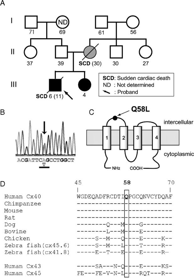
Figure 1.
GJA5 mutation identified in a family with the clinical diagnosis of PFHB1. A: Family pedigree. Genetically affected and unaffected individuals shown with closed and open symbols, respectively. Hatched circle: Proband's mother not genotyped; clinical data suggest she was a de novo mutation carrier. Number below each symbol: age at registration or age of sudden death (parenthesis). B: Sequence electropherogram of exon 2 GJA5 of proband. Arrow indicates heterozygous missense mutation of leucine (CTG) for glutamine-58 (CAG). C: Cx40 predicted membrane topology indicating position Q58 in first extracellular loop. D: Sequence alignment of human Cx40 and its homologues (residues 45-70). Notice also conservation in human Cx43 and Cx45. Dashes indicate residues identical with the top sequence.

