
Figure 5.
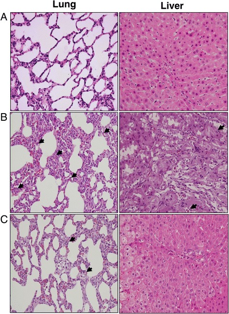
Histopathological studies of lung and liver. Light microscopy showed lung and liver sections of rats in groups of (A) sham operation, (B) cecal ligation and puncture (CLP), and (C) CLP plus Levosimendan administration. Sections were stained with haematoxylin and eosin. Arrows represent polymorphonuclear neutrophil infiltration in lung and liver. Each is shown at 400 × (original magnification).