Figure 5.

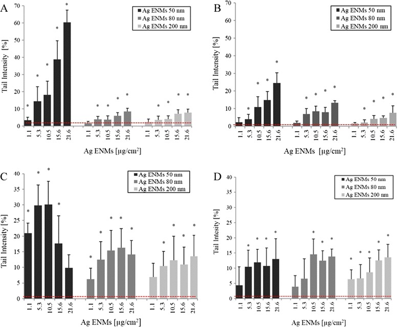
Level of DNA damage – strand breaks (A, B) and oxidised DNA lesions expressed as NET FPG ( C, D) in A549 cells exposed to different concentrations of Ag ENMs ( μg/ cm 2 ) with sizes 50, 80, 200 nm for 2 h ( A, C) and 24 h (B, D). NET FPG was estimated as FPG-sensitive sites minus strand breaks, representing altered purines. Horizontal lines represent level of strands breaks (SBs)/oxidised bases (NET FPG) in untreated cells (A: 1.36 ± 0.84; B: 0.76 ± 0.5; C: 1.99 ± 1.7; D: 3.3 ± 1.56% tail DNA). The data are expressed as mean ± SD of three independent experiments. *significant (p < 0.05) difference from the unexposed control. Hydrogen peroxide (50 μM, 5 min in PBS), a positive control (SBs) gave 38.72 ± 6.1% tail DNA. Photosensitiser Ro19-8022 (1 μM in PBS, 5 min, on ice), a positive control for oxidised DNA lesions (NET FPG) gave 33.2 ± 10.96% tail DNA.
