
Fig. 1. Schematic of molecular counting technology.
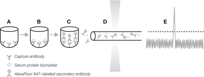
Serum protein biomarkers were quantified with molecular counting technology in a 10-μL volume using a 384-microwell immunoassay format. (A), Attached to the surface of each microwell are capture antibodies specific for an individual biomarker. (B), Upon addition of serum sample, biomarkers are bound to the capture antibodies, followed by the addition and binding of AlexaFluor 647-labeled secondary antibody (C). (D), The fluorescence-labeled antibody complexes are chemically released from each well and pumped through a capillary flow system for detection of laser-induced fluorescence. (E), Photons emitted from AlexaFluor 647–labeled antibody molecules are distinguished from background levels so that each signal represents a molecular counting event.