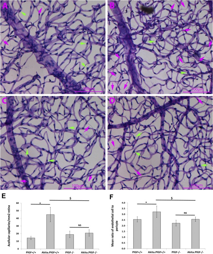
Figure 3.
Decreased capillary degeneration and pericyte loss in the retinas of Akita.PlGF−/− mice. Six-month-old diabetic male mice (∼5-month diabetes duration) and nondiabetic littermate male mice were used for elastase digestion. A–D: Representative examples of isolated retinal vasculature for PlGF−/− (A), Akita.PlGF−/− (B), WT (C), and Akita (D) mice strains. Purple arrows indicate acellular capillaries. Green arrows indicate pericytes. E: Quantification of acellular capillaries. Results were expressed as mean ± SD number of acellular capillaries/mm2. F: Pericyte quantification. The results were expressed as the ratio of ECs to pericyte (n = 6). *P < 0.05 vs. nondiabetic WT control; $P < 0.05 vs. Akita diabetic mice; NS, not significant.

