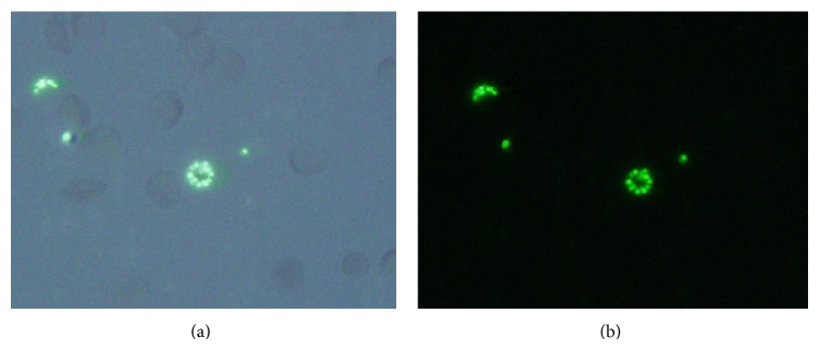
Figure 1.

P. falciparum infected RBCs were stained with SYBR Green I and were then examined using a microscope with (a) DAPI and (b) fluorescence filter. Photographs indicated fluorescent images of parasites at schizont (center) and merozoite stages (outer).
