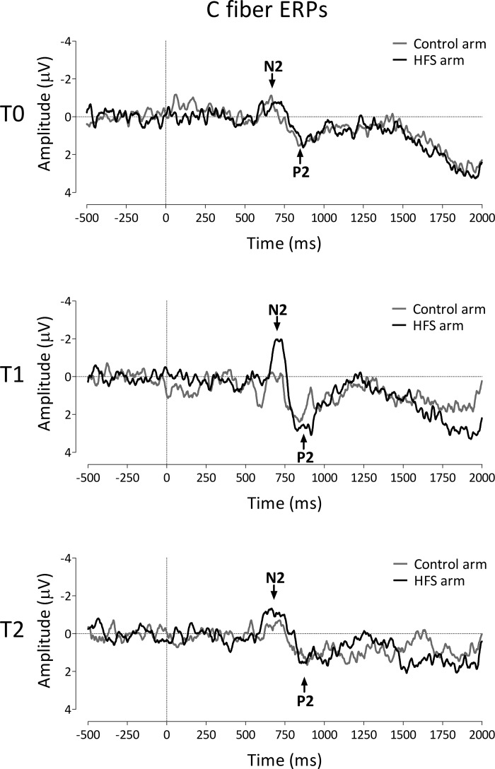
Fig. 4.
Effect of HFS on the event-related brain potentials (ERPs) elicited by the thermal laser stimuli selectively activating C fibers. The waveforms show the group-level average ERP waveforms of the signals measured from Cz vs. average reference, before HFS (T0), 20 min after HFS (T1), and 45 min after HFS (T2), following stimulation of the HFS-treated arm (black) and the control arm (grey). Note the increase of the N2 wave at the treated arm at T1.

