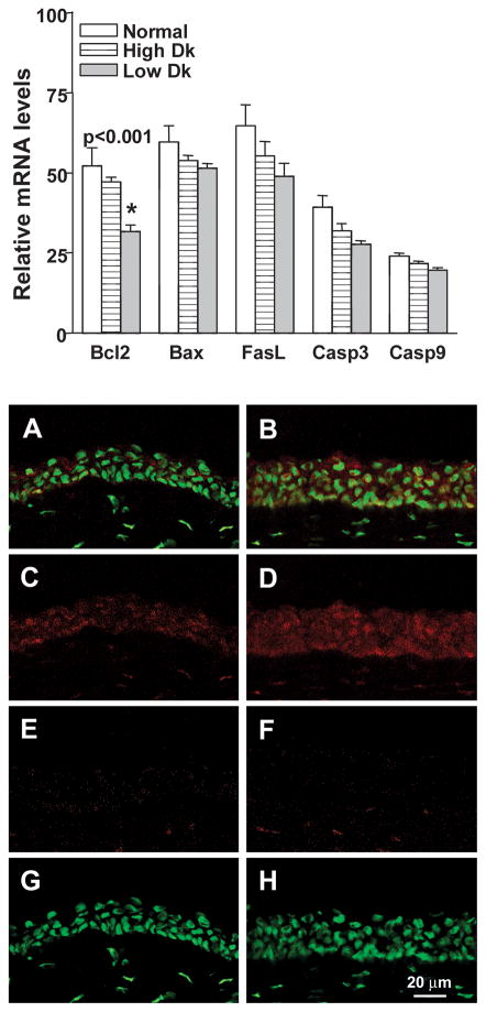
Figure 5.
(Top) Real time RT-PCR at 48 hours after bacterial challenge in normal, and uninfected high and low Dk wearing eyes. mRNA levels of Bcl-2 were significantly decreased in the infected low Dk wearing corneas when compared with levels in the normal or challenged but uninfected high Dk CL wearing corneas. No differences among the three groups were seen for Bax, FasL, caspase 3 or caspase 9. (Bottom A–H). More cells in the epithelium appeared to be stained positively for Bcl-2 in the high (B,D) vs low Dk (A,C) CL wearing cornea. (E, F) Control eyes incubated in the absence of the primary antibody showed no staining in the epithelium, but some non-specific keratocyte staining was observed. (G, H) SYTOX GREEN nuclear labeling alone is shown. Magnification= 220X; bar= 20μm.

