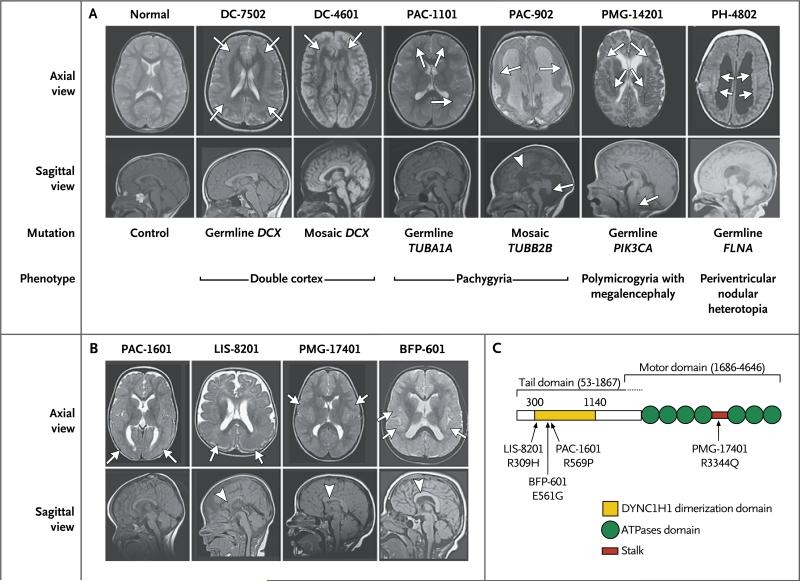
Figure 3. MRI Features of Brain Malformations Associated with Germline and Mosaic Variants, and Positions of Variants within the DYNC1H1 Protein.
Panel A shows axial and midline sagittal MRI scans of the head of a person without neurologic abnormalities; two persons with the double-cortex syndrome (white arrows), associated with germline variants in one (Participant DC-7502) and a mosaic variant in the other (Participant DC-4601); two participants with pachygyria (white arrows), associated with germline variants in one (Participant PAC-1101) and a mosaic variant in the other (Participant PAC-902); a person with polymicrogyria (white arrows) and megalencephaly associated with a germline variant (Participant PMG-14201); and a person with periventricular nodular heterotopia (white arrows) associated with germline variants (Participant PH-4802). In addition, the sagittal image in Participant PAC-902 shows dysgenesis of the corpus callo- sum (arrowhead) and hypoplastic cerebellar vermis (white arrow), and the sagittal image of Participant 14201 shows cerebellar tonsillar herniation (white arrow) due to mass effect from the enlarged cerebral hemispheres. In Panel B, axial and midline sagittal MRI scans in the four persons with de novo mutations in DYNC1H1 show posterior-predominant pachygyria (white arrows in axial views in Partici- pants PAC-1601 and LIS-8201), thick, irregular cortex in the perisylvian region (white arrows in scans from Participants PMG-17401 and BFP-601), and a mildly dysmorphic corpus callosum (arrowheads in sagittal views in Participants LIS-8201, PMG-1704, and BFP-601). Panel C is a schematic representation of the DYNC1H1 protein, showing the positions of the alterations (black arrows) detected in our study.

