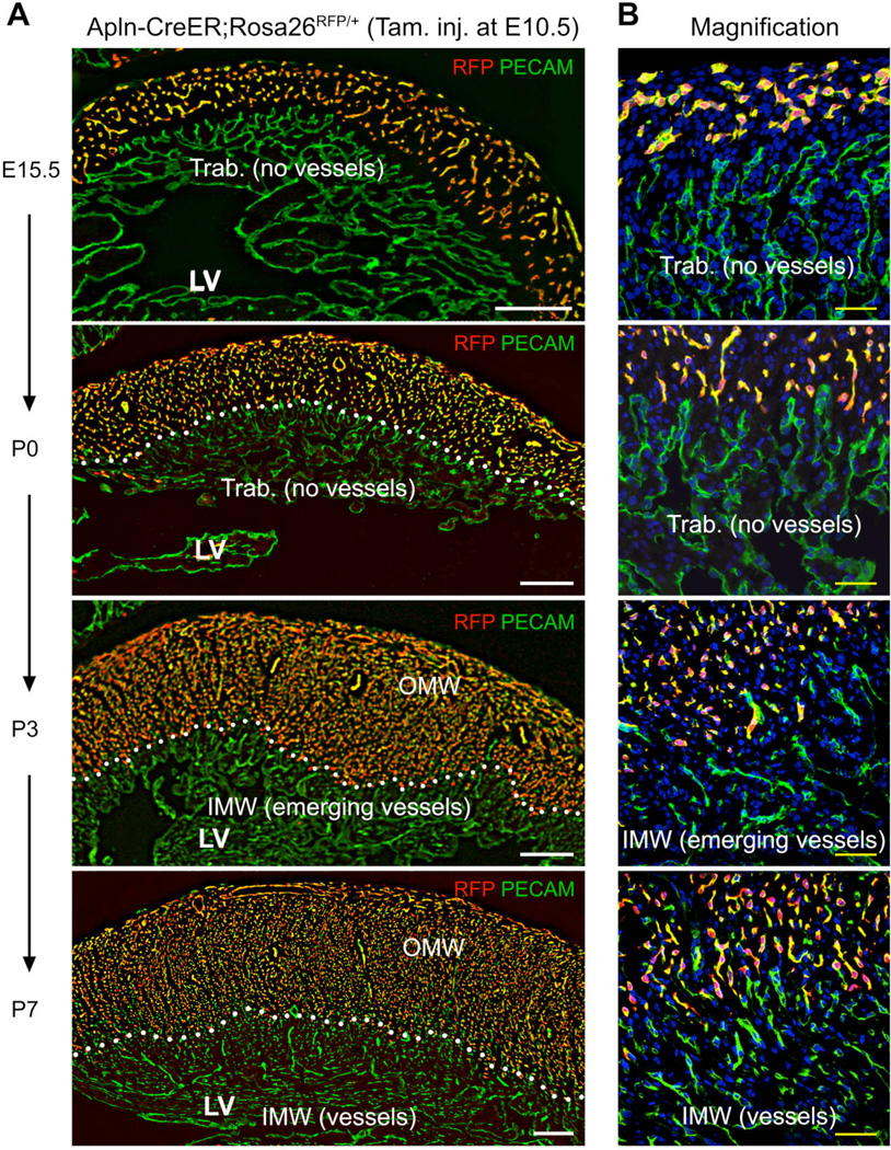
Fig. 1. Derivatives of embryonic coronary vessels do not expand into the inner myocardial wall of the neonatal heart.
(A, B) Embryonic coronary vessels were RFP-labeled by tamoxifen treatment of Apln-CreER;Rosa26RFP/+ embryos at E10.5. In the neonatal heart, these VECs, derived from embryonic coronary vessels, occupied the outer portion of the compact myocardium, defined as the Outer Myocardial Wall (OMW). The remaining, RFP negative portion of the compact myocardium was defined as the Inner Myocardial Wall (IMW). White dotted lines highlight the inner border of left ventricular myocardium occupied by embryonically labeled vessels. From P0 to P7, Trabecular myocardium (Trab.) transformed into compact myocardium where new coronary vessels arise. White bar = 200 µm; yellow bar = 50 µm. LV, left ventricle.

