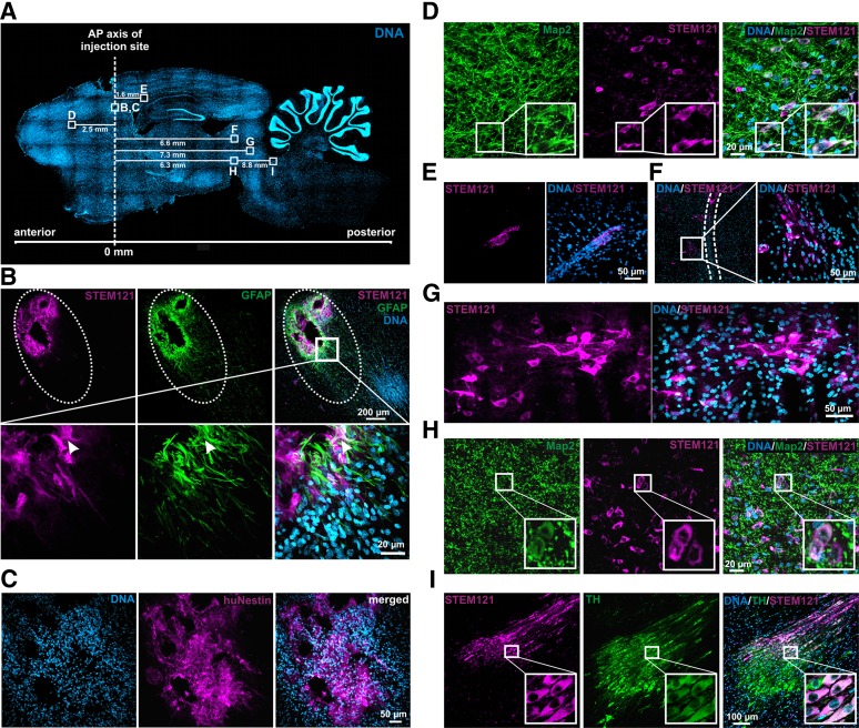
Figure 6.
Adult human neural crest-derived stem cells derived from the inferior turbinate (ITSCs) survive and integrate into parkinsonian rat brains. Immunohistochemical analysis of sagittal 6-hydroxydopamine (6-OHDA) rat brain sections at 12 weeks after transplantation. (A): Whole-brain section stained with DAPI (4′,6-diamidino-2-phenylindole). Dotted line indicates AP axis of injection site. Squares depicting regions of interest with appropriate migration distances. (B): Cluster of STEM121+ cells were directly located at the striatal injection site (upper panel). Dotted ellipse illustrates the injection channel. GFAP+ astrocytes were recruited to the injury site. Magnified view: arrowhead indicates STEM121+/GFAP+ coexpressing cell (lower panel). (C): ITSCs remaining at the injection site kept their stem cell characteristics and expressed the human-specific neural crest stem cell marker nestin. (D): STEM121+/Map2+ ITSCs were shown to be located within the corpus callosum directly above the striatum. (E): At 12 weeks after transplantation, several STEM121+ cells remained within the corpus callosum 1.6 mm posterior to the injection site. (F): In addition, STEM121+ ITSCs were located adjacent to the injection channel of the 6-OHDA lesion, indicated by dotted lines (G), and 7.3 mm posterior to the striatal injection site within the midbrain. (H): STEM121+/Map2+ ITSCs integrated directly above the lesioned SN. (I): Stem121+ ITSCs located in the locus ceruleus showed TH coexpression at the protein level, indicating their differentiation into DA neurons. Abbreviations: AP, anteroposterior; hNestin, human-specific neural crest stem cell marker nestin; TH, tyrosine hydroxylase.

