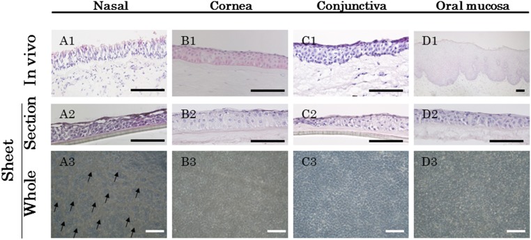
Figure 1.
Histological examination of the cultivated nasal mucosal epithelial cell sheet. Light micrographs showing cross-sections of nasal (A1, A2), corneal (B1, B2), conjunctival (C1, C2), and oral (D1, D2) epithelial cells (ECs), both native tissue and in the cultured sheet stained with hematoxylin and eosin. Phase contrast images showing a confluent primary culture of nasal (A3), corneal (B3), conjunctival (C3), and oral (D3) ECs after 2 weeks in culture. Arrows indicate cells with a goblet-like appearance (A3). Scale bars = 100 μm.

