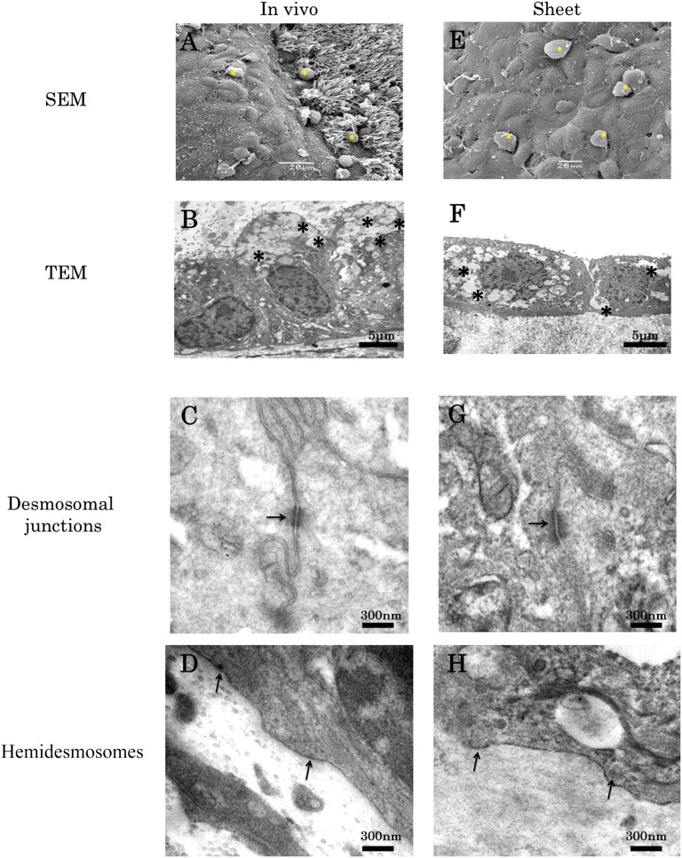
Figure 2.
Ultrastructural examination of the cultivated nasal mucosal epithelial cell sheet (CNMES). Scanning electron microscopy examination of native nasal mucosa (A) and the CNMES (E). Yellow asterisks indicate blobs of mucus. Transmission electron microscopy examination of native nasal mucosa (B, C, D) and the CNMES (F, G, H). Asterisks indicate mucus-filled vesicles (B, F). Arrows indicate desmosomes (C, G) and hemidesmosomes (D, H). Abbreviations: SEM, scanning electron microscopy; TEM, transmission electron microscopy.

