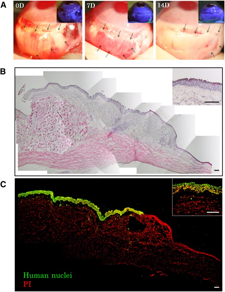
Figure 5.
Xenotransplantation of a human cultivated nasal mucosal epithelial cell sheet. (A): Representative slit-lamp photographs of a rabbit taken immediately after transplantation, 7 days after transplantation, and 14 days after transplantation, with and without fluorescein. Arrows indicate the transplanted cell sheet. (B, C): Hematoxylin and eosin staining (B) and immunofluorescence (C) of anti-human nuclei at the transplanted conjunctival area. Scale bars = 100 μm. Abbreviations: D, day(s); PI, propidium iodide.

