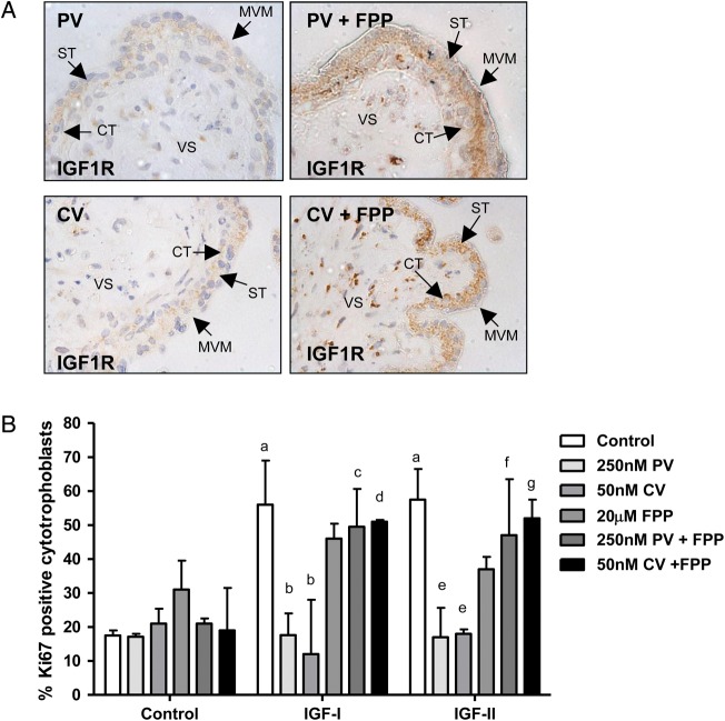
Figure 4.
The effect of 3-Hydroxy-3-methylglutaryl co-enzyme A (HMG-CoA) reductase inhibitors on insulin-like growth factor 1 receptor (IGF1R) cell surface expression and IGF-induced proliferation can be attenuated by co-incubation with glycosylation inhibitors. First trimester placental explants (n = 5) were incubated in the absence or presence of cerivastatin (CV; 50 nM) or pravastatin (PV; 250 nM) alone or in combination with farensyl pyrophosphate (FPP; 20 μM) for 24 h before the addition of IGF-I (10 nM) or IGF-II (10 nM) and culture for a further 24 h. (A) IGF1R localization was analysed by immunohistochemistry using an IGF1R-specific antibody. Arrows indicate microvillous membrane (MVM), syncytiotrophoblast (ST), cytotrophoblast (CT) and villous stroma. Pravastatin and cerivastatin do not disrupt IGF1R expression in the presence of FPP. Each image is representative of staining observed in three individual placentas. Scale bars on images represent 50 μM. (B) Proliferation was assessed by using immunohistochemistry to determine the number of Ki67-positive cytotrophoblast as a percentage of total cytotrophoblast and data are presented as median and range of five independent experiments. A Kruskal–Wallis test followed by a Dunn's post hoc test was used to assess significance (P < 0.05; n = 5) a = different from control, untreated tissue; b = different from IGF-I treated tissue; c = different from tissue treated with pravastatin and IGF-I; d = different from tissue treated with cerivastatin and IGF-I ; e = different from IGF-II treated tissue; f = different from tissue treated with pravastatin and IGF-II; g = different from tissue treated with cerivastatin and IGF-II.

