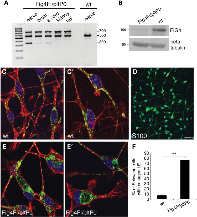
Figure 4.
P0-Cre-mediated recombination efficiency in the Fig4Floxed/plt, P0-Cre model. (A) PCR analysis on genomic DNA from Fig4Floxed/plt, P0-Cre mice and controls. A 350-bp recombination band was detected in the nerve of mutants but not in wild type. The 350-bp faint band in the brain and spinal cord indicates recombination in cranial and spinal nerves of brain and spinal cord, respectively. (B) Western blot analysis of nerve homogenates at P30 indicates decreased Fig4 expression in sciatic nerves of Fig4Floxed/plt, P0-Cre mice. (C–F) Purified Schwann cells from Fig4Floxed/plt, P0-Cre mouse nerves at P3 indicate the presence of enlarged LE/LY-LAMP1 positive (green in C, C′, E, E′; red is phalloidin; blue is DAPI) in mutant cells, a feature of Fig4 loss and PtdIns(3,5)P2 decrease. (D) S100 staining marks Schwann cells in the culture. Bar in (D) is 20 µm for (C, C′, E, E′) and 50 µm for (D).

