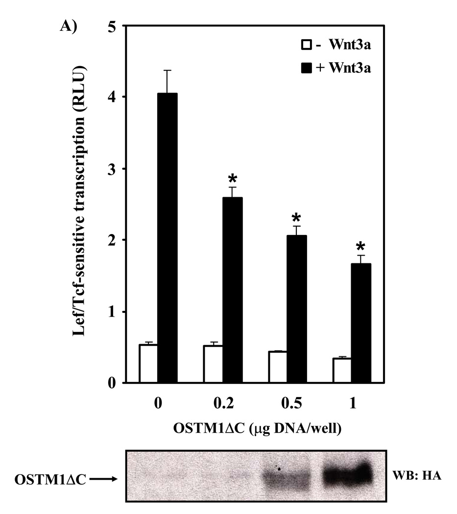
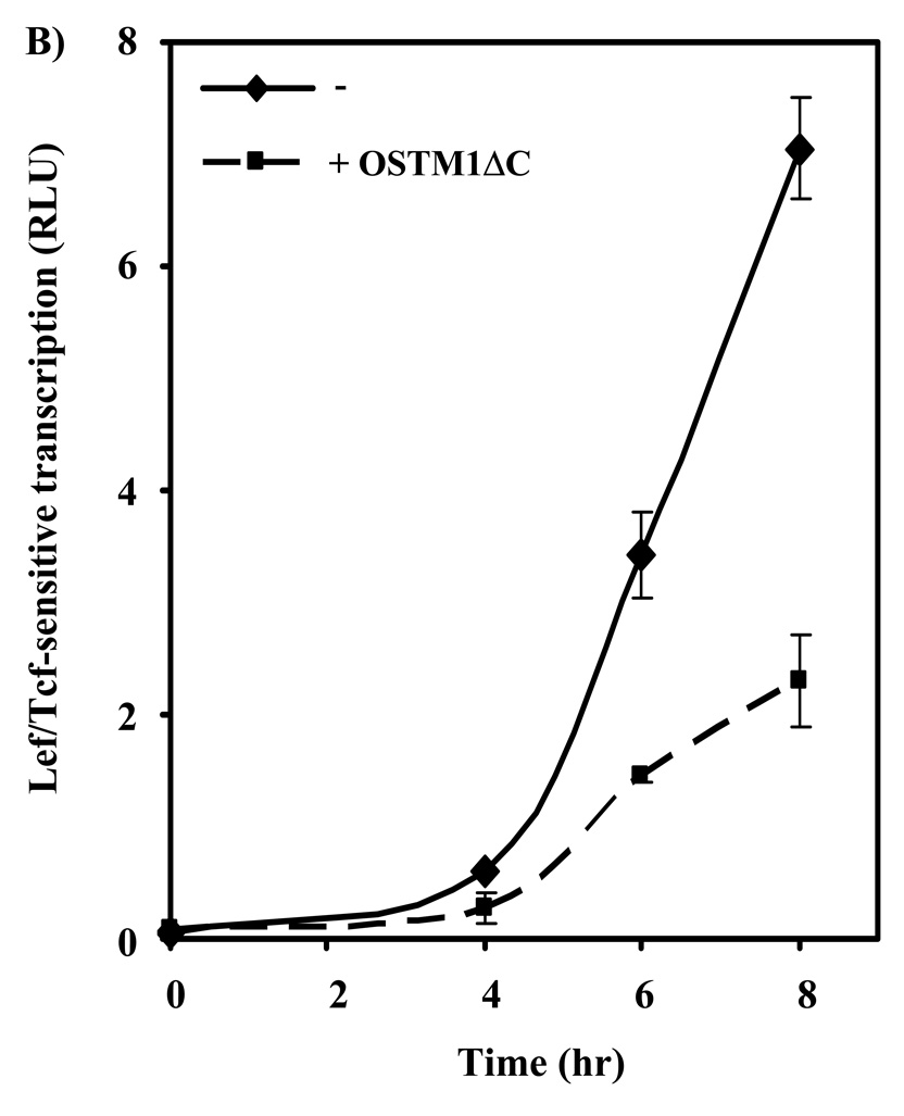
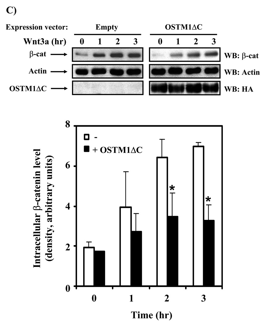
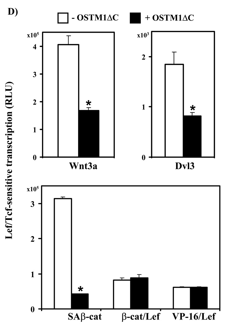
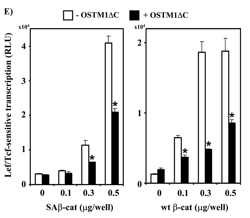
Figure 4. OSTM1ΔC mutant attenuates Wnt/β-catenin signaling.
(A) F9 cells transiently transfected with Fz1, M50 and either empty vector or the indicated amounts of OSTM1ΔC were treated with Wnt3a for 7 hr, then collected and subjected to the luciferase assay. * denotes P < 0.05 for the difference between wild-type, Wnt3a-treated cells and those expressing OSTM1ΔC and Wnt3a-treated for the individual doses. Lower panel: Western blot analysis was used to measure the expression of transiently transfected HA-tagged OSTM1ΔC. (B) F9 cells transiently transfected with Fz1, M50 and either empty vector or OSTM1ΔC were treated with Wnt3a for the indicated lengths of time, then collected and subjected to luciferase assay. (C) F9 cells transiently transfected with Fz1 and either empty vector or OSTM1ΔC were treated with Wnt3a for the indicated lengths of time. Ly sates were collected and treated with concanavalin A to separate cytosolic from membrane-associated β-catenin. Cleared samples were subjected to SDS-PAGE on an 11% acrylamide gel, transferred to nitrocellulose and probed with an antibody against β-catenin. The results displayed are from a single experiment representative of more than three independent tests. Lower panel: Bands from multiple gels were quantified by densitometry. * denotes P < 0.05 for the difference between wild-type cells and those expressing OSTM1ΔC for the individual time points. (D) F9 cells transiently transfected with Fz1, M50 and the indicated constructs (or treated with Wnt3a for 7 hr) were collected and subjected to luciferase assay. * denotes P < 0.05 for the difference between Wnt/β-catenin pathway activated cells and those expressing OSTM1ΔC. (E) F9 cells transiently transfected with Fz1, M50, OSTM1ΔC and the indicated amounts of SAβ-catenin or wild-type β-catenin were collected and subjected to luciferase assay. * denotes P < 0.05 for the difference between cells transfected with SAβ-catenin or wild-type β-catenin and an empty vector and those transfected with SAβ-catenin or wild-type β-catenin and OSTM1ΔC.

