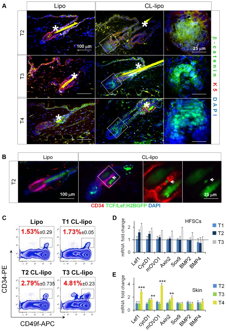
Figure 4. Reduction of skin macrophages is associated with activation of β-catenin/Wnt signaling.
(A) Immunofluorescence analysis of β-catenin (green) and K5 (red) in backskin sections of mice treated with CL-lipo and Lipo controls; n = 4. *Hair shaft autofluorescence. Bar = 25 µm. (B) Immunofluorescence analysis of CD34 (red) and H2B-GFP signal (green) in T2 backskin sections of TCF/Lef:H2B-GFP transgenic mice treated with CL-lipo and Lipo controls; n = 3. Arrows point to GFP positive cells. Bar = 25 µm. The gating strategy is shown in Figure S11 B. (C) FACS analysis of single cell suspensions of CD34+CD49f+ HF-SCs (gated) isolated from backskin of mice treated with CL-lipo or Lipo controls at specified time points; n = 2–4. (D) Relative mRNA expression of canonical Wnt/β-catenin target genes and BMP signaling genes in HF-SCs isolated as indicated in (B); n = 2–4. (E) Relative mRNA expression of canonical Wnt/β-catenin target genes and BMP signaling genes in total back-skin samples after treatment with CL-lipo compared to Lipo controls; n = 6. Note: n refers to the number of mice, per point per condition. *p≤0.05. All data used to generate the histograms can be found in Data S1.

