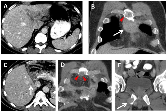
Figure 8. A 57 year old male with BAP1 mutated intrahepatic CCA.
Baseline (A) axial and (B) coronal contrast-enhanced CT images demonstrate (A) a 6.9×5.3 cm liver mass involving segments I, II, and IV, and (B) a 5.6 cm sternal metastasis (arrow). Note the sternal metastasis does not extend to the level of the right 3rd rib (arrowhead). Extended left hepatectomy and resection of the sternal metastasis was performed. (C, D) Seven weeks later, contrast-enhanced CT images show a new metastasis in the remnant liver (arrow), and a new 2 cm recurrence (arrowheads) involving the sternum adjacent to the right 3rd rib. (E) Three months later, there is a new 2.2 cm metastasis (arrow) involving the right inferior pubic ramus.

