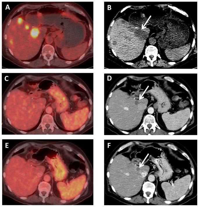
Figure 11. A 67 year old male with BRAF mutated intrahepatic CCA, who had progressed on conventional chemotherapy.
Axial (A) fused PET-CT and (B) unenhanced CT images from a PET scan demonstrate FDG avidity of multiple liver metastases. After 8 weeks of BRAF inhibitor therapy, axial (C) fused PET-CT and (D) contrast-enhanced CT images demonstrate lack of FDG avidity and decreased size of liver metastases, e.g., the dominant lesion adjacent to the IVC (arrow) decreased from 3.7 cm to 1.6 cm. After 16 weeks of therapy, axial (E) fused PET-CT and (F) contrast-enhanced CT images demonstrate continued lack of FDG avidity and further decreased size of liver metastases, e.g., the dominant lesion (arrow) now measures 1.3 cm.

