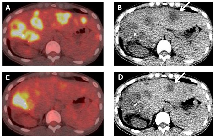
Figure 12. A 37 year old female with c-MET amplified CCA, who had progressed on conventional chemotherapy.
At baseline, axial (A) fused PET-CT and (B) unenhanced CT images demonstrate multiple FDG avid liver metastases, in this patient who is status post right hepatectomy. After 4 weeks of therapy, (C) axial fused PET-CT and (D) unenhanced CT images show decreased FDG avidity and size of metastases. A representative segment II mass (arrows) has decreased from 2.6 cm to 2.1 cm.

