
Figure 1. CD90 expression in normal human pancreas.
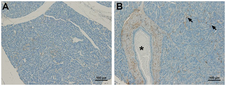
(A) A representative image of normal pancreas showed negative expression of CD90. (B) Minimal CD90 expression was observed on the connective tissues (arrows). The fibroblasts surrounding the pancreatic main duct (asterisk) showed weak CD90 expression. No expression of CD90 was observed on ducts, acini, islets, and blood vessels. Scale bars = 100 µm.