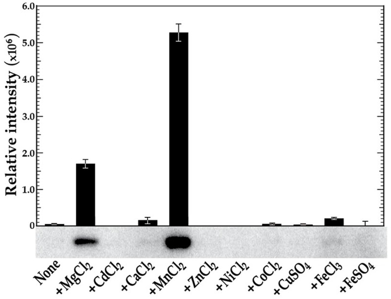
Figure 1. in vitro phosphorylation of VicK in the presence of various metal cations.
VicK (1 µM) was incubated in 100 mM Tris-HCl, pH 7.5 containing 1 mM of the designated cations and 0.10 µM [γ-32P] ATP at room temperature for 15 minutes. The relative autophosphorylation of VicK was quantified using Image Quant 5.0 software (Molecular Dynamics) and is represented by the histogram above the scanned gel. The gels shown are representative of at least three independent experiments. Error bars represent ± std. errors of the average phosphorylation values derived from at least 3 independent experiments.

