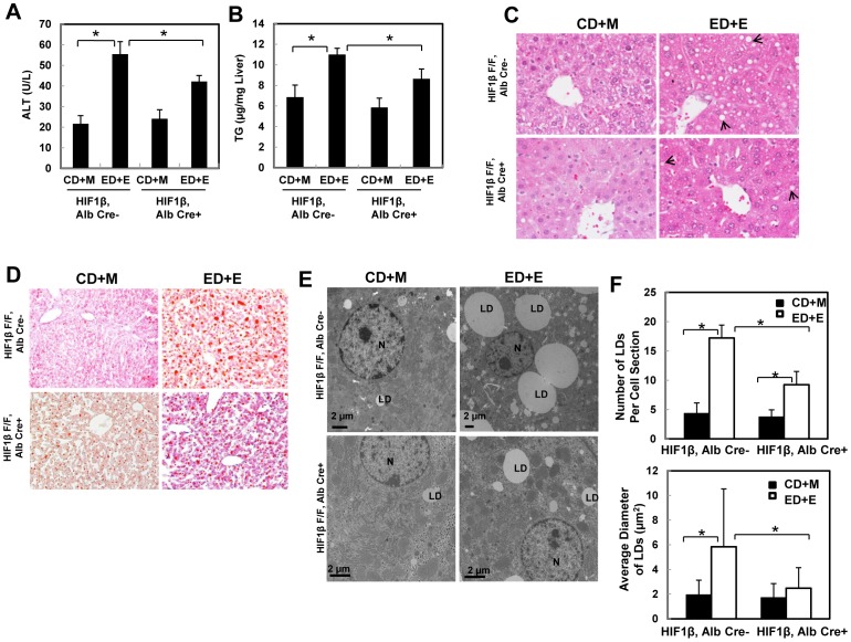
Figure 4. Hepatocyte-specific HIF-1β knockout mice were resistant to steatosis and liver injury from the Gao binge model.
Age matched male wild type and hepatocyte-specific HIF-1β knockout mice were subjected to Gao-binge treatment. Serum ALT (A) and hepatic TG (B) were measured. Data are presented as means ± SE (n = 3–8). * p<0.05. One way ANOVA with Scheffé's post hoc test. Representative photographs of H &E staining (C) and Oil O Red staining are shown (D). Arrows: hepatic lipid droplets. Representative EM images are shown in (E). M: Mitochondria; N: Nuclei; LD: lipid droplet; Bar: 500 nm. The number and size (average diameter) of LDs per cell section was quantified (F), and data are presented as means ± SE (more than 20 cell sections and 80 LDs). * p<0.05. One way anova analysis with Scheffé's post hoc test.

